
Mini C-Arm types are standard in most outpatient surgery facilities and are used for emergency medicine, orthopedic procedures, and pain management. C-Arms are equipped with radiographic capabilities and are used for intraoperative imaging during the listed procedures. It’s non-invasive equipment, making it safe after and during screening procedures.
Orthoscan DI Mini C-arm
Mobile DI is a mini C-Arm ideal for off-site extremity imaging needs for those who sustain injuries. The portable fluoroscopic and X-ray device provides multiple connectivity options for electronic management records and offers excellent digital image quality. The mid-mount monitor arm provides increased horizontal and vertical motion, allowing for optimal display while standing or sitting. The main benefits of this device include the following:
- Image flexibility: Mobile DI can access shoulder images besides foot and weight-bearing knee views. The Orthoscan DI mini C-Arm can be mounted on the accessory cart or placed on a table, enabling more effortless movement between rooms. The accessory cart provides C-Arm rotation for knee and shoulder views.
- Portable and lightweight: Orthoscan mobile DI guarantees ease of movement between satellite clinics, exam rooms, emergency departments, military units, athletic team venues, and urgent care centers. Mobile DI offers versatility for orthopedic imaging and has the smallest footprint available for mini C-Arms.
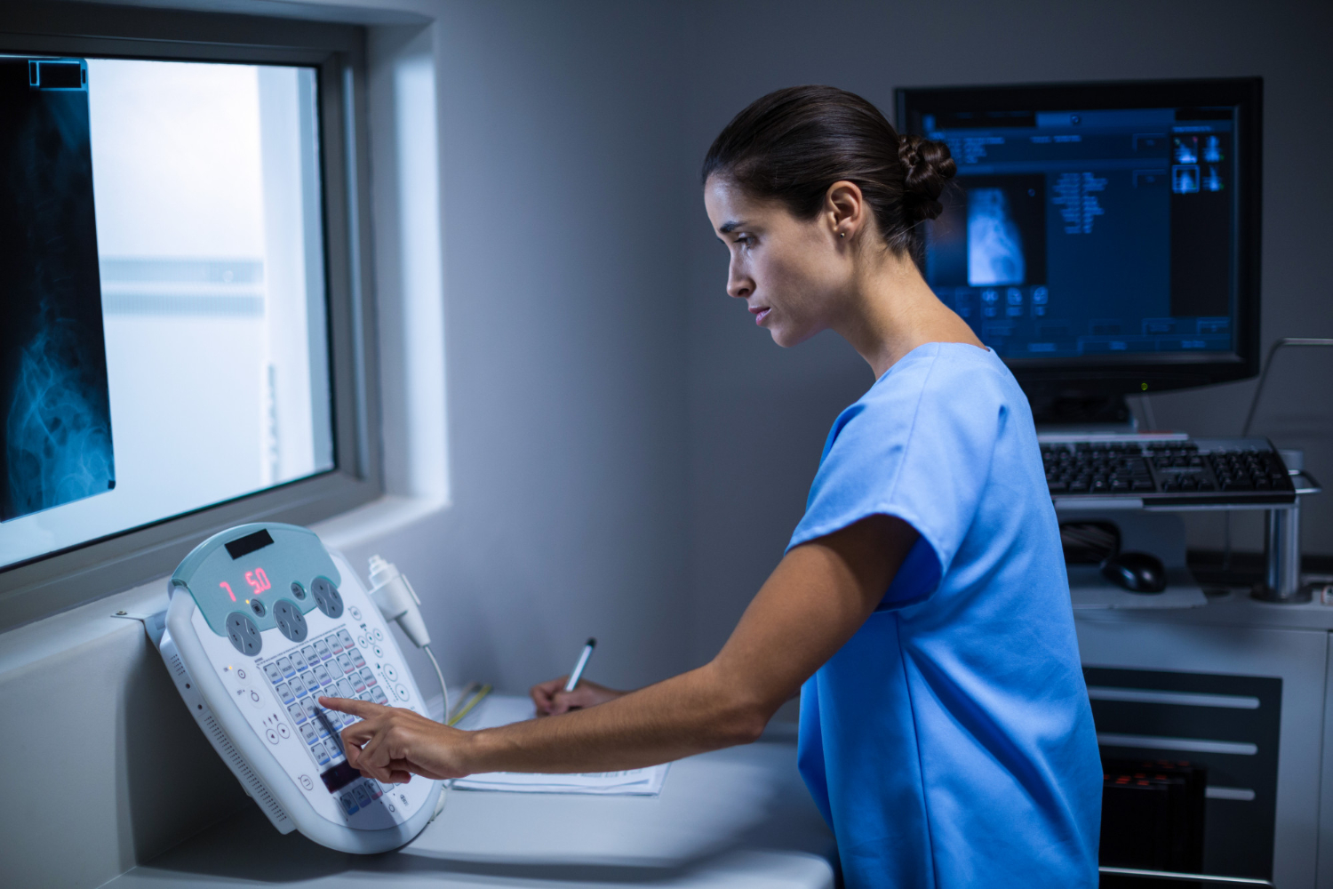
Components of a C-arm System
A portable C-Arm system contains three parts: an imaging system, an X-ray generator, and a workstation unit.
Imaging System
The C-Arm has a powerful imaging system capable of performing multiple movements in one procedure. The much-needed advantage is essential during various surgical procedures, namely urology, ortho, and cardiology. The entire system is light and compact in weight to facilitate multiple positioning and a wide range of movement. The system remains firm when mounted to minimize the possibility of misalignment during the procedure.
X-ray Generator
The generator is positioned in the same frame where the C-Arm gets mounted. The workstation unit controls it directly, allowing the operator to modify the operation in real-time. Even with a surge in X-ray power, there’s total flexibility in imaging, and the risk involved is almost null due to minimal exposure time.
Workstation Unit
The overall operation of the C-Arm is done with the assistance of a workstation unit. It contains various handles facilitating positioning and movements, switches controlling light exposure and power, a cable hanger, and controls for fluoro and radiographic settings. The workstation unit also contains advanced image enhancement software, which reduces noises, hard disk and writers, zoom control, monitors, and a brake pedal.
Fluoroscopy
Fluoroscopy is an imaging procedure that utilizes continuous pulses in an X-ray beam to obtain real-time footage of tissues and organs inside our body. The primary difference between fluoroscopy and X-rays is that the former provides continuous real-time images while the latter captures snapshots in a single moment. Healthcare professionals utilize the imaging technique for diagnosis and insertion of cardiac catheterization, intravenous injections, and catheter placement. The fluoroscopy technology offers extremities that yield real-time high-resolution images. It enables surgeons to perform correct procedures through continuous snapshots of the happenings inside the tissue.
Healthcare professionals must take good care of the C-Arm machine to maintain its efficiency. Make sure to clean the filters and wheels and wipe the external parts regularly. Request professional assistance if there are any issues with your machine.